
Nagaveni NB 1,2,3
1Consultant Pedodontist, “Garike Dental Care”, Davangere, Karnataka, India
2Consultant Pedodontist, Karnataka ENT Hospital and Research Centre, Chitradurga, Karnataka, India.
3Professor, Department of Pediatric and Preventive Dentistry, College of Dental Sciences, Davangere, Karnataka, India
Corresponding author: Nagaveni NB, Consultant Pedodontist, “Garike Dental Care”, Davangere, Karnataka, India.
Received: November 17, 2023
Accepted: November 28, 2023
Published: December 13, 2023
Citation: Nagaveni NB. (2023) “Pink Tooth of Mummery or Odontoclastoma in Pediatric Patients: A Case Series”. Dental Science and Innovative Research, 2(1); DOI: http;//doi.org/03.2023/1.1002.
Copyright: © 2023 Nagaveni NB. This is an open access article distributed under the Creative Commons Attribution License, which permits unrestricted use, distribution, and reproduction in any medium, provided the original work is properly cited.
The purpose of this article is to show case few cases of ‘Pink tooth of Mummery’ also called as ‘Odontoclastoma’ or ‘Internal Resorption’ which were diagnosed in primary dentition. Although it is a normal physiologic process, knowledge about its occurrence is highly essential among clinicians for proper diagnosis.
Introduction
The present article aims to show case few cases of ‘Pink tooth of Mummery’ which is also called by various synonyms as ‘Odontoclastoma’ or ‘Internal resorption’ or ‘Chronic perforating hyperplasia of pulp’ and ‘Internal granuloma’ which were encountered in pediatric patients (Figure 1). Table 1 shows elaborative description of ‘Pink tooth of Mummery’ cases.
Table 1: Detailed description of cases found with “Pink tooth of Mummery”
|
Case No. |
Age (in years) |
Gender |
Tooth found with pink spot |
Type of dentition |
|
1.[Figure 1, A] |
12 |
Male |
Mandibular right second molar |
Primary dentition |
|
2. [Figure 1, B] |
9 |
Male |
Maxillary right lateral incisor |
Primary dentition |
|
3. [Figure 1, C] |
10 |
Female |
Maxillary right second molar |
Primary dentition |
|
4. [Figure 1, D] |
11 |
Female |
Maxillary left lateral incisor |
Primary dentition |
|
5. [Figure 1, E] |
9 |
Male |
Maxillary left lateral incisor |
Primary dentition |
‘Pink tooth of Mummery’ was found in five patients. Age of the patient ranged from 9 to 12 years. Among five cases, there were three male and two females. All five cases were found in primary dentition and the teeth affected by pink spot phenomenon were mandibular right second molar, maxillary right lateral incisor, maxillary right second molar, maxillary left lateral incisor and maxillary left lateral incisor (Figure 1, A-E).
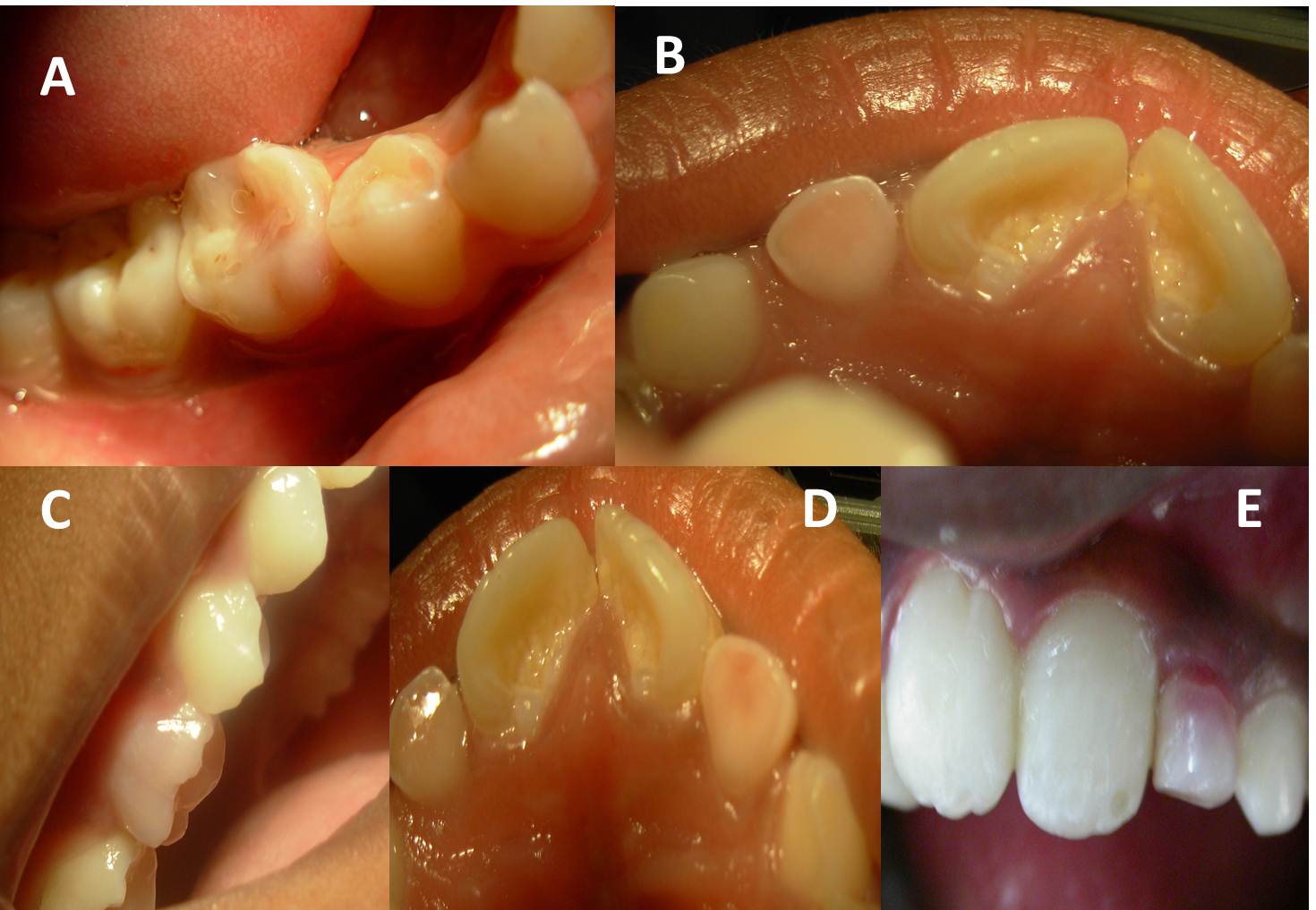
Figure 1: Photograph showing ‘Pink tooth of Mummery’ in primary teeth
A famous anatomist James Howard Mummery in 1920, called it as ‘Pink tooth of Mummery’ due to the presence of a pink discoloration exhibited in the crown portion of the tooth. Pink discoloration occurs because of the loss of dentin creating a large pulp space which then allows more blood vessels to fill the area and leads to pinkish hue to the crown structure, whereas, scientist Fothergil named it as ‘pink spot’ [1]. In 1920, Mummery extensively studied about pink spots and later published detail literature on it. Pritchard showed histologically that internal resorption is comparable to a granuloma of the pulp. Although internal resorption is a rare occurrence, any injury to the pulp tissue, like physical trauma or caries-related pulpitis can lead to internal resorption in the tooth [2]. This condition is mostly found either in the mid or apical area which is noticed by accidentally following radiographic examination, or by the presence of clinical sign of a ‘pink spot’ on the crown. The pulp within the tooth show partial or complete necrosis and in such cases of actively progressing lesion, the tooth may be partially vital and may present symptoms of typical pulpitis [2].
Root resorption is the loss of dental hard tissues as a result of clastic activities and it may be due to either physiologic or pathologic phenomenon. Root resorption in the primary dentition is a normal physiologic process except when the resorption occurs prematurely. The initiating factors involved in physiologic root resorption in the primary dentition are not completely understood, although the process appears to be regulated by cytokines and transcription factors that are similar to those involved in bone remodeling [3,4].
Compared to bone, as it undergoes continuous physiologic remodeling throughout life, root resorption of permanent teeth does not occur naturally and is invariably inflammatory in nature. Therefore, root resorption in the permanent dentition is a pathologic event, and if not treated in time, this might result in the premature loss of the affected teeth. In differential diagnosis, other conditions where pink discoloration of the teeth is seen should be evaluated. They are Lepromatous leprosy, hypothermia, trimipramine intoxication and pneumonia [4]. Pertaining to permanent teeth, occurrence of pink tooth has some clinical implications. It is considered to be an important aspect of forensic science, where in the phenomenon of post-mortem pink teeth were seen in cases of drowning, strangling and suffocation. The possibility of occurrence of pinkish appearance of the tooth in such cases is because of the marked congestion in the head region, leading to congestion within the pulp followed by hemorrhage and diffusion within the pulp chamber [4,5].

Open Access By Aditum Open Access Journals id licensed under Creative Commons Attribution 4.0 International License. Based On a Work at aditum.org